Ophthalmoscopy (also called fundoscopy) is an exam your doctor, optometrist, or ophthalmologist uses to look into the back of your eye. With it, they can see the retina (which senses light and images), the optic disk (where the optic nerve takes the information to the brain), and blood vessels.
What can a ophthalmoscope detect?
It is used to detect and evaluate symptoms of retinal detachment or eye diseases such as glaucoma. Ophthalmoscopy may also be done if you have signs or symptoms of high blood pressure, diabetes, or other diseases that affect the blood vessels.
What part of eye Cannot be seen with ophthalmoscope?
The optic disc, also called the blind spot, is where the axons of the ganglion cells leave the retina to form the optic nerve. It is called the blind spot because there are no rod or cone receptors in this part of the retina and we can not see objects that are imaged on this part of the retina.
What structures can be viewed through an ophthalmoscope?
The retinal structures viewed through the ophthalmoscope are the optic disc, the retinal vessels, the general background, and the macula.What pathological conditions can be detected with an Ophthalmoscopic exam?
Fundoscopic / Ophthalmoscopic Exam. Visualization of the retina can provide lots of information about a medical diagnosis. These diagnoses include high blood pressure, diabetes, increased pressure in the brain and infections like endocarditis.
Which parts of the ophthalmoscope are present on the front of the ophthalmoscope head quizlet?
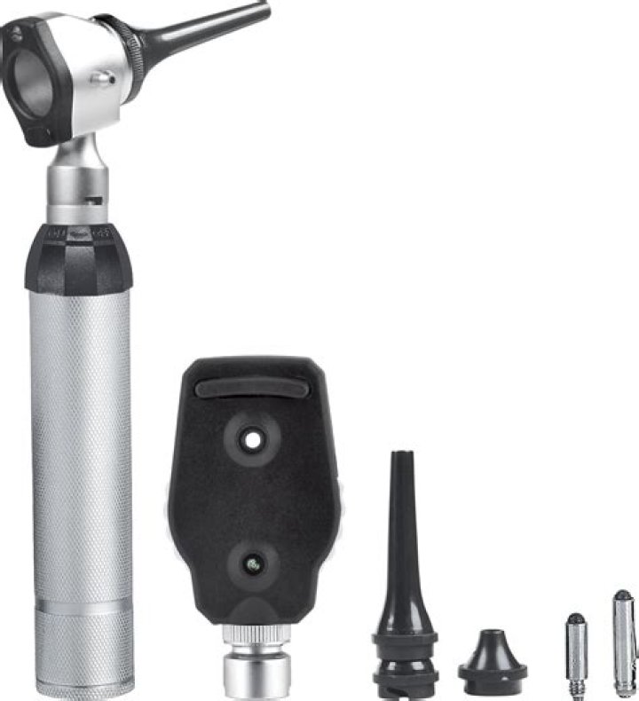
The head of the ophthalmoscope consists of five different parts: viewing aperture, aperture selector, mirror window, lens selector, and lens indicator. The mirror window is present on the front, because it enables the examiner to look through the pupil at the background of the eye.
What does a Retinoscope do?
Retinoscopy (also called skiascopy) is a technique to objectively determine the refractive error of the eye (farsighted, nearsighted, astigmatism) and the need for glasses. The test can be quick, easy, reliably accurate and requires minimal cooperation from the patient.
Are otoscope and ophthalmoscope the same?
Otoscopes are used in ear examinations. A doctor uses these instruments to look into the ear canal to look at the ear drum. … An ophthalmoscope is and instrument that lets the doctor to look into the back of your eye known as the fundus.What is Echo ophthalmoscope?
D009887. Ophthalmoscopy, also called funduscopy, is a test that allows a health professional to see inside the fundus of the eye and other structures using an ophthalmoscope (or funduscope). It is done as part of an eye examination and may be done as part of a routine physical examination.
When using the ophthalmoscope it is best to?Place your left hand on the patient’s head and place your thumb on their eyebrow. Hold the ophthalmoscope about 6 inches from the eye and 15 degrees to the right of the patient. Find the red reflex. Move in closer, staying nasally until you see the optic nerve.
Article first time published onWhat is green light on ophthalmoscope?
Red-free light is little used, although it is an elementary method. Green-light ophthalmoscopy, with its short wavelength, enhances some fundus and vitreous structures and may make the examination of pathologic conditions (premacular pathology, vascular abnormalities, etc.)
How do you find the optic disc with an ophthalmoscope?
Begin at arm’s length by shining the ophthalmoscope light into the patient’s pupil (you will then see the red reflex). Follow this reflex until your forehead rests on your thumb—you should immediately see the optic disc.
What is the direct ophthalmoscope?
A direct ophthalmoscope, or simply an ophthalmoscope, is a hand-held optical instrument used to inspect the fundus or back of the eye.
Which type of mirror is used in ophthalmoscope to examine throat and ear?
Concave mirrors are used in optical instruments such as Ophthalmoscope.
How do you clean an ophthalmoscope?
To clean the exterior of your ophthalmoscope, prepare a 70% isopropyl alcohol solution and soak a lint-free cloth. Wipe down the exterior of the ophthalmoscope head and handle, cleaning all external surface areas. Care should be taken to prevent excess liquid from seeping into the components.
What is plane mirror retinoscope?
Copeland streak retinoscope: All the way up, plane mirror is in position with a wide streak.As it is lowered gradually , the streak decreases in width. And widens again.At the lowest adjustment the streak is again at its maximum width but with concave mirror effect.
What does it mean if my child has a Anisometropia?
Anisometropia means that the two eyes have a different refractive power (glasses prescription), so there is unequal focus between the two eyes.
What is streak retinoscope?
of Optometry And According to the Dictionary Visual Science; 5th Edition,pg:267, streak retinoscope is a retinoscope which projects into the patient’s eye an oblong streak which can be adjusted in width and rotated in various meridians.
Which instrument is used to examine both the ear and the nose?
An otoscope is an instrument which is used to look into the ear canal. The ear speculum (a cone-shaped viewing piece of the otoscope) is slowly inserted into the ear canal while looking into the otoscope. The speculum is angled slightly toward the person’s nose to follow the canal.
Which equipment measures the range of motion of a shoulder joint?
A goniometer is a device used in physical therapy to measure a joint’s range of motion (ROM).
Which equipment would the nurse use to assess a patient's blood pressure select all that apply?
Device Summary Stethoscope or doppler and a blood pressure cuff with a mercury or aneroid sphygmomanometer, or automated oscillometric blood pressure measuring device.
How does ophthalmoscope look like?
An ophthalmoscope is an instrument that has a light and several small lenses on it. Your eye doctor can look through the lenses to examine your eye. They may ask you to look in certain directions as they conduct the examination.
What does a fundus photo show?
Fundus photographs are ocular documentation that record the appearance of a patient’s retina. Optometrists, ophthalmologists, orthoptists and other trained medical professionals use fundus photography for monitoring the progression of certain eye condition/diseases.
Who uses an otoscope?
An otoscope or auriscope is a medical device which is used to look into the ears. Health care providers use otoscopes to screen for illness during regular check-ups and also to investigate ear symptoms. An otoscope potentially gives a view of the ear canal and tympanic membrane or eardrum.
Can you use otoscope for eyes?
The otoscope is a valuable tool beyond its primary role as an examination tool for detecting ear problems. It can also be used for transillumination, dermatologic observation, examination of the eye and body orifices other than the ear, as a pump, as a light source, in veterinary medicine, and in non-medical tasks.
How much is an ophthalmoscope?
How Much Does ophthalmoscope cost? The prices of some instruments can be as low as 60 USD or less, while the newest, high-tier systems may cost as much as 9000 USD. Well-known brands of ophthalmoscope in the United States are Heine, Keeler and Welch Allyn.
How are ophthalmoscopes used in animals?
In using the direct ophthalmoscope, place the lens dial at 0 setting and hold the ophthalmoscope at 20 inches from the patient’s eye. Observe the pupil and tapetal reflex. Opacities within the lens can be visualized at this time.
What is indirect ophthalmoscope?
The binocular indirect ophthalmoscope, or indirect ophthalmoscope, is an optical instrument worn on the examiner’s head, and sometimes attached to spectacles, that is used to inspect the fundus or back of the eye. It produces an stereoscopic image with between 2x and 5x magnification.
How do you use ophthalmoscope for red reflex?
Hold the ophthalmoscope close to your eyes. Encourage the child to look at the light source and direct the light at the child’s eyes individually and together. You should see an equal and bright red reflex from each pupil. Pay attention to the colour and brightness of the red reflex.
Who invented ophthalmoscope?
Results: Hermann von Helmholtz, German physician and physicist, presented and published his invention of the ophthalmoscope in 1851. Albrecht von Graefe was the first to use ophthalmoscope routinely. He said: ‘Helmholtz has opened a new world to us’.
Where was the ophthalmoscope invented?
Xavier Galezowski of Paris, France, who had invented his tubular indirect ophthalmoscope in 1862, designed a very different ophthalmoscope (Figure 11) 20 years later in 1882. This used a single Rekoss disc but with 2 concentric rings of lenses mounted within it.