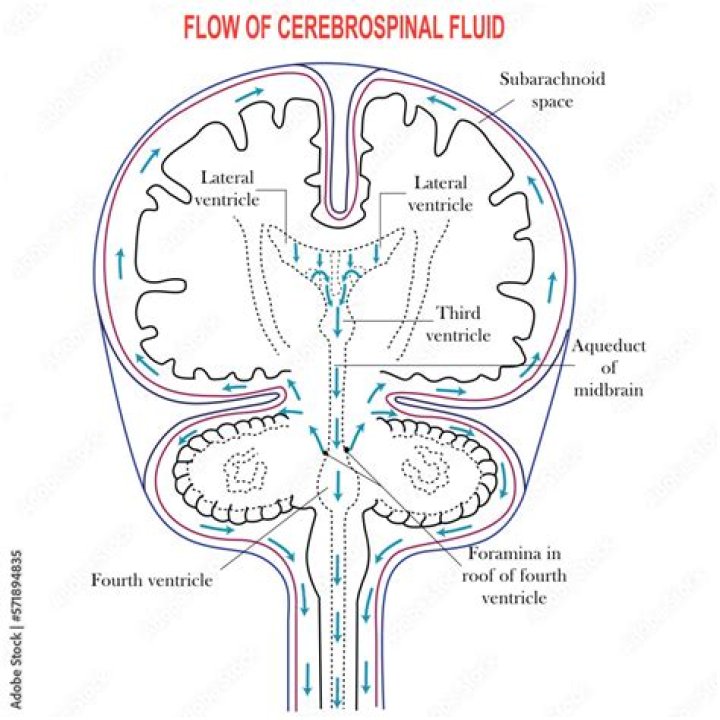
A series of interconnected, fluid-filled cavities are found within the brain. These cavities are the ventricles of the brain, and the fluid is cerebrospinal fluid (CSF).
What is the name of the fluid filled cavity of midbrain?
The cavity of the mesencephalon forms the cerebral aqueduct. The dilation of the neural canal within the rhombencephalon forms the fourth ventricle.
What fluid filled space is between the pons and cerebellum?
The fourth ventricle is between the pons and the cerebellum and within the superior portion of the medulla oblongata. The fourth ventricle communicates with the central canal of the spinal cord.
What are the large fluid filled spaces within the brain called?
The large fluid-filled spaces within the brain are called ventricles. There are four of them, and they are filled with cerebrospinal fluid. The formation of cerebrospinal fluid occurs in the choroid plexuses by removing fluid from the blood.What ventricle is in the midbrain?
The third ventricle is continuous caudally with the cerebral aqueduct, which runs though the midbrain. At its caudal end, the aqueduct opens into the fourth ventricle, a larger space in the dorsal pons and medulla. The fourth ventricle narrows caudally to form the central canal of the spinal cord.
Where is CSF made?
CSF formation. Most CSF is formed in the cerebral ventricles. Possible sites of origin include the choroid plexus, the ependyma, and the parenchyma[2]. Anatomically, choroid plexus tissue is floating in the cerebrospinal fluid of the lateral, third, and fourth ventricles.
What is forebrain?
forebrain, also called prosencephalon, region of the developing vertebrate brain; it includes the telencephalon, which contains the cerebral hemispheres, and, under these, the diencephalon, which contains the thalamus, hypothalamus, epithalamus, and subthalamus.
Is cerebrospinal fluid found in the subdural space?
The classic view has been that a so-called subdural space is located between the arachnoid and dura and that subdural hematomas or hygromas are the result of blood or cerebrospinal fluid accumulating in this (preexisting) space.When does the process of myelination complete?
The corticospinal tract starts to myelinate at 36 weeks gestation and myelination is completed by the end of the 2nd year of life. Myelination of the corticospinal tract begins at the proximal portion of the axon and the shortest axons are the first to myelinate.
Does pia mater contain CSF?Function. In conjunction with the other meningeal membranes, pia mater functions to cover and protect the central nervous system (CNS), to protect the blood vessels and enclose the venous sinuses near the CNS, to contain the cerebrospinal fluid (CSF) and to form partitions with the skull.
Article first time published onWhat part of the spinal cord is filled with cerebrospinal fluid CSF?
The space under the arachnoid, the subarachnoid space, is filled with cerebrospinal fluid and contains blood vessels. The pia mater is the innermost layer of meninges.
What is the slit like fluid filled cavity that separates the right and left halves of the diencephalon?
The third ventricle is one of the four connected ventricles of the ventricular system within the mammalian brain. It is a slit-like cavity formed in the diencephalon between the two thalami, in the midline between the right and left lateral ventricles, and is filled with cerebrospinal fluid (CSF).
What does the midbrain develop into?
Unlike the other two vesicles – the forebrain and hindbrain – the midbrain does not develop further subdivision for the remainder of neural development. It does not split into other brain areas. while the forebrain, for example, divides into the telencephalon and the diencephalon.
What cerebrospinal fluid does?
Also called CSF. Cerebrospinal fluid (CSF, shown in blue) is made by tissue that lines the ventricles (hollow spaces) in the brain. It flows in and around the brain and spinal cord to help cushion them from injury and provide nutrients.
Which ventricle is in the brainstem anterior to the cerebellum?
The fourth ventricle is a diamond-shaped cavity located dorsal to the pons and upper medulla oblongata and anterior to the cerebellum (Fig. 1.13).
Where does fluid flow from the cerebral aqueduct?
The CSF passes from the lateral ventricles to the third ventricle through the interventricular foramen (of Monro). From the third ventricle, the CSF flows through the cerebral aqueduct (of Sylvius) to the fourth ventricle.
Is the thalamus in the midbrain or forebrain?
The forebrain, midbrain and hindbrain make up the three major parts of the brain. The structures in the forebrain include the cerebrum, thalamus, hypothalamus, pituitary gland, limbic system, and the olfactory bulb. The midbrain consists of various cranial nerve nuclei, tectum, tegmentum, colliculi, and crura cerebi.
Is the hypothalamus part of the midbrain?
According to the typical division of the brain into the forebrain, midbrain, and hindbrain, the hypothalamus is a part of the forebrain. It is considered to be a part of the diencephalon. … Anteriorly, it extends up to the optic chiasma and posteriorly it is continuous with the tegmentum of midbrain.
Is temporal lobe in midbrain?
Midbrain (Mesencephalon) There are three parts to the midbrain: the colliculi, the tegmentum, and the cerebral peduncles. The colliculi processes visual and auditory signals before they are relayed to the occipital and temporal lobes.
Is cerebrospinal fluid found in the epidural space?
The epidural space is the area between the dura mater (a membrane) and the vertebral wall, containing fat and small blood vessels. The space is located just outside the dural sac which surrounds the nerve roots and is filled with cerebrospinal fluid.
What is spiral cord?
A column of nerve tissue that runs from the base of the skull down the center of the back. It is covered by three thin layers of protective tissue called membranes. The spinal cord and membranes are surrounded by the vertebrae (back bones).
How does myelination occur in the CNS?
The myelination of axons throughout the nervous system is one such crucial maturation process. In the central nervous system (CNS), glial cells called oligodendrocytes extend many processes into their surrounding environment, which concentrically wrap membrane around axons to form myelin sheaths.
What is the function of the dendrites in a neuron?
Nerve cells (neurons) have extensive processes called dendrites. These occupy a large surface area of a neuron. They receive many signals from other neurons and contain specialized proteins that receive, process, and transfer these to the cell body.
What is myelinated nerve fibers?
Myelinated retinal nerve fiber layers (MRNF) are retinal nerve fibers anterior to the lamina cribrosa that, unlike normal retinal nerve fibers, have a myelin sheath. Clinically, they appear to be gray-white well-demarcated patches with frayed borders on the anterior surface of the neurosensory retina.
What is CSF fluid made of?
Cerebrospinal fluid (CSF) is a clear, colourless ultrafiltrate of plasma with low protein content and few cells. The CSF is mainly produced by the choroid plexus, but also by the ependymal lining cells of the brain’s ventricular system.
What is EPI PIA and PIA glia?
ADVERTISEMENTS: The pia mater consists of epi-pia which lines the arachnoid andpia-glia (pia intima) which covers the brain surface. Both epi-pia and pia-glia are connected by strands of pial cells across the subarachnoid space. No sub-pial space intervenes between the pia mater and the underlying brain surface.
Is pia mater innervated?
The vessels of the cortex, the pia mater and the dura mater all receive their sensory fibers, for the most part, from the trigeminal ganglion. Fibers coming from other cranial nerves, from the medulla oblongata and the upper cervical region also contribute to this innervation.
Is the cauda equina made of pia mater?
The cauda equina exists within the lumbar cistern, a gap between the arachnoid membrane and the pia matter of the spinal cord, called the subarachnoid space.
Which of the following spaces around the spinal cord is filled with cerebrospinal fluid CSF )? Quizlet?
subarachnoid space: the space between the pia and arachnoid mater of the brain and spinal cord that contains cerebrospinal fluid (CSF).
What are the spaces found in the spinal cord?
The pia mater is the innermost protective layer and is tightly associated with the surface of the spinal cord. The space between the arachnoid and pia maters is called the subarachnoid space and is where the CSF is located.
Which membrane is found lining the brain and spinal cord?
Three layers of membranes known as meninges protect the brain and spinal cord. The delicate inner layer is the pia mater. The middle layer is the arachnoid, a web-like structure filled with fluid that cushions the brain. The tough outer layer is called the dura mater.