Nevus comedonicus is an uncommon skin abnormality first described in 1895 by Kofmann who used the term “comedo nevus.” It comprises of groups of pits filled with black keratinous plugs resembling blackheads, with inflammatory acne lesions developing later.
How do you treat nevus comedonicus?
As nevus comedonicus is an asymptomatic benign condition, no treatment is required. Surgical excision of small lesions can be curative and should be considered in consultation with a dermatologic/plastic surgeon. Incomplete excision may result in recurrence of the lesion. Laser surgery has been tried.
How rare is nevus comedonicus?
Nevus comedonicus is rare, accounting for only 1% of epidermal nevi. Only about 200 cases have been reported to date. In a retrospective examination of 417,511 pediatric patients in Mexico City, 443 patients were found to have epidermal nevi. Five of these patients were found to have nevus comedonicus.
What does nevus comedonicus look like?
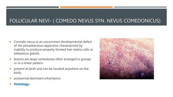
Nevus comedonicus is characterized by closely arranged, grouped, often linear papules that have at their center dilated follicular openings with keratinous plugs resembling comedones. Cysts, abscesses, fistulas, and scars develop in about half the cases, which have been described as “inflammatory” nevus comedonicus.How many people have nevus comedonicus?
Nevus comedonicus is a rare problem with an estimated occurrence of 1 case in every 45,000–100,000 individuals [2, 6].
What causes nevus of Ota?
Cause. Nevus of Ota is caused by the entrapment of melanocytes in the upper third of the dermis. It is found only on the face, most commonly unilaterally, rarely bilaterally and involves the first two branches of the trigeminal nerve. The sclera is involved in two-thirds of cases (causing an increased risk of glaucoma) …
Can congenital moles be cancerous?
Most congenital nevi do not cause health problems and may just require monitoring for the development of skin cancer. Large congenital nevi are associated with an increased risk of developing an aggressive form of skin cancer known as melanoma.
What is epidermal nevus syndrome?
Epidermal nevus syndromes (ENSs) are a group of rare complex disorders characterized by the presence of skin lesions known as epidermal nevi associated with additional extra-cutaneous abnormalities, most often affecting the brain, eye and skeletal systems.Is nevus Comedonicus real?
Nevus comedonicus syndrome is a rare syndrome with extracutaneous manifestations. Commonly involved sites for comedo nevus include face, neck, upper arm, chest and abdomen in a grouped, band-like lesions, or linear pattern along lines of Blaschko.
How are comedones formed?Comedones arise when cells lining the sebaceous duct proliferate (cornification), and there is increased sebum production. A comedo is formed by the debris blocking the sebaceous duct and hair follicle. It is now known that comedones also involve inflammation (see causes of acne).
Article first time published onWhat is nevus Anemicus?
Nevus anemicus is a congenital vascular anomaly that presents clinically as a hypopigmented macule or patch, as shown below. The lesional pallor is due to a localized hypersensitivity to catecholamines with resultant vasoconstriction. Nevus anemicus is an uncommon disorder and was first described by Vorner in 1906.
What is Favre Racouchot?
Favre-Racouchot syndrome is a disorder consisting of multiple open and closed comedones in the presence of actinically damaged skin. The disease was originally described in 1932 by Favre and reviewed in detail by Favre and Racouchot in 1951.
How common is nevus Depigmentosus?
The term nevus depigmentosus, however, is a misnomer, because the lesion is hypopigmented but not depigmented. The reported prevalence of nevus depigmentosus varies from 0.4% to 3%.
How much does it cost to remove nevus?
Typical costs: Removal of a mole typically costs about $150 to $400. It varies from doctor to doctor and by which technique is used.
Is a compound nevus benign or malignant?
Compound naevi are considered to be benign neoplasms of melanocytes if they arise in later life. Compound naevi arise from a flat (junctional) naevus that exists earlier in life and may have a raised central portion of deeper pigmentation with surrounding tan-brown macular pigmentation.
How do you treat epidermis nevus?
The treatment of choice for small epidermal nevi is surgical excision. Superficial means of removal frequently result in recurrence. Aggressive approaches may be more successful, but also carry a higher risk of postoperative scarring.
What does a congenital nevus look like?
A congenital nevus looks like a round or oval-shaped patch of colored skin and is usually raised. They can be either a single color or multi-colored. They can vary in size from a tiny spot to something that covers a large part of your body. In some cases, they might have hair growing out of them.
When should I be concerned about a mole on my child?
If a mole bleeds without reason, however, it should be checked. A mole that looks like an open sore is also worrisome. Bleeding or a break in the skin can be a sign of melanoma. Bottom line: If your child has a mole that starts to bleed or looks like an open sore, a dermatologist should examine the mole.
Do congenital nevi grow?
If they are seen at birth or develop during the first 1-2 years of life they are called congenital melanocytic nevi. While most of these moles are small, some may be very large. Most of these will grow as your child grows.
Can nevus be removed?
Small- and medium-sized nevi are generally harmless but can be removed for aesthetic reasons. Removal of a small nevus consists of a simple excision, cutting out the affected area and stitching the surrounding skin back together. Removing a large nevus is a much more daunting process.
How do you lighten nevus of Ota?
Laser treatments are the most effective corrective approach to nevus of Ota, although they must be repeated more than once, with multiple approaches and applications. The laser treatments work to destroy the melanocytes that cause the bluish hyperpigmentation, with the goal of returning the skin to its natural pigment.
Is Nevus of Ota congenital?
It comprises dermal melanocytes, presumably arising due to the dermal arrest of cells migrating from the neural crest. Nevus of Ota is 3-5 times more common in women than men, and is classified into congenital type, appearing soon after birth, and acquired type, appearing during or after puberty.
How do you close a dilated pore of Winer?
Your healthcare provider will close large dilated pores of Winer with stitches after removing the contents of the pore. Small dilated pores of Winer, similar to the size of a traditional blackhead, should close on their own after squeezing the contents of the pore out with tweezers.
What is inflammatory linear verrucous epidermal nevus?
Inflammatory linear verrucous epidermal nevus (ILVEN) is a type of skin overgrowth, called epidermal nevus. It is characterized by skin colored, brown, or reddish, wart-like papules (nevi). The nevi join to form patches or plaques that often follow a pattern on the skin known as the “lines of Blaschko“.
Is linear epidermal nevus hereditary?
It usually occurs alone; however, rarely ILVEN can be associated with other symptoms as part of an epidermal nevus syndrome. Rarely, ILVEN can become cancerous (for example, basal cell carcinoma or squamous cell carcinoma). ILVEN is caused by a genetic change that occurs after conception ( somatic mutation ).
Is a nevus a mole?
Most people continue to develop new moles until about age 40. In older people, common moles tend to fade away. Another name for a mole is a nevus. The plural is nevi.
Is nevus sebaceous hereditary?
Linear nevus sebaceous syndrome (LNSS) is not inherited . All cases reported have been sporadic, occurring by chance in people with no family history of the condition. While LNSS is caused by genetic mutations , these mutations occur after fertilization in the affected person.
What is junctional nevus?
A junctional nevus is a mole that occurs between two layers of skin. This image shows two junctional nevi that appear as uniformly brown small macules, round in shape with smooth regular borders. Color Atlas & Synopsis of Pediatric Dermatology.
Why am I getting so many closed comedones?
But if you have a lot of them, and they’re fairly constant, you probably have a type of acne called comedonal acne. Closed comedones are really common during the tween and teen years. This is because when you are a pre-teen or a teenager, the skin’s sebaceous glands (also known as oil glands) speed up production.
How long do closed comedones last?
How long does a comedone last? A closed comedone will usually last 1 to 3 weeks but can last longer at times.
What happens inside a comedone?
Comedones are small flesh-colored, white, or dark bumps on your skin. They are a type of acne blemish, caused by plugs of oil and dead skin that become stuck in the openings (follicles) that enclose the roots of your hair. A single bump is called a comedo .