Indication Statement. The Titanium Trochanteric Fixation Nail (TFN) is intended to treat stable and unstable fractures of the proximal femur including pertrochanteric fractures, intertrochanteric fractures, basal neck fractures, and combinations thereof.
What is TFN A?
The TFN-ADVANCED Proximal Femoral Nailing System (TFNA) is designed to solve a wide range of unmet needs for surgeons, OR staff, and administrators. More than five years in the making, this system offers advancement in hip fracture treatment.
What is Cephalomedullary nail?
Cephalomedullary nailing is the surgical stabilization of the fracture with an intramedullary device usually inserted through the piriformis fossa, the tip or lateral greater trochanter, or the medial greater trochanter.
What is proximal femoral nailing?
The proximal femoral nail (PFN) is an osteosynthetic implant designed to treat proximal femoral fractures in the trochanter area with a closed intramedullary fixation method.What is Gamma nail surgery?
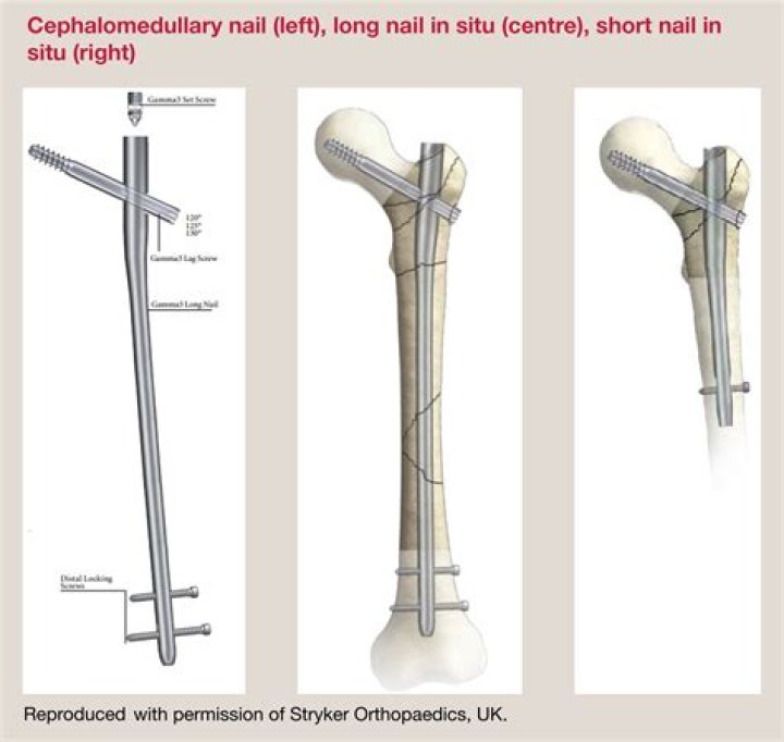
Gamma is a comprehensive intramedullary nailing system for the treatment of a wide range of proximal femur fractures as well as associated femoral shaft fractures. The system is the result of over 25 years of continuous innovation and clinical history.
What is subtrochanteric femur fracture?
Subtrochanteric femoral fractures are fractures of the proximal femur that may extend proximally into the piriformis fossa or distally into the isthmus of the femur. The proximal extension of the fracture varies and may include fracture patterns combined with intertrochanteric and femoral neck fractures.
What does TFNA mean?
AcronymDefinitionTFNATesticular Fine Needle Aspiration (urology)TFNATransthoracic Fine Needle Aspiration (pulmonology)TFNATesla Foundation of North America
What is PFNA2?
PFNA2 (Proximal femoral nail antirotation for Asia) has been developed especially for Asian patients. The treatment of proximal femoral fractures in geriatric osteoporotic patients continues to be a challenge in orthopedic trauma.Should intramedullary nails be removed?
Intramedullary nail removal is safe. Patients with anterior knee pain should be told that their pain may persist and that knee pain may even arise.
What is intramedullary nailing?An intramedullary nail is a metal rod that is inserted into the medullary cavity of a bone and across the fracture in order to provide a solid support for the fractured bone. Intramedullary nailing is currently considered the “gold standard” for treatment of femoral shaft fractures (Rudloff 2009).
Article first time published onIs Cephalomedullary nail the same as intramedullary nail?
This new class of intramedullary nail, the so-called “cephalomedullary nail” includes the Long Gamma Nail (LGN), the Trochanteric Femoral Nail (TFN), and the Intertroch/Subtroch Nail (ITST).
What is a hip CMN?
Cephalomedullary nails (CMN) are commonly used for the treatment of intertrochanteric (IT) hip fractures. Total hip arthroplasty (THA) is commonly used as a salvage procedure for failed IT hip fractures that progress to post-traumatic arthritis.
How does a Cephalomedullary nail work?
The nail features a small proximal section that is designed to minimize the amount of bone that must be removed for nail insertion. A lag screw is placed through the nail into the femoral head to secure the nail in place proximally and help control the different segments of the bone while healing occurs.
Is a gamma nail an ORIF?
References. Normal femur anatomy. Femur with plate and screws. Femur with intramedullary rod and screw.
What are Gamma nails made of?
Material: Titanium alloy with anodized type II surface treatment. Locking in the distal part of the oblong hole creates a dynamic locking mechanism − requires only one screw (see Fig. 2).
What does a gamma nail look like?
The Gamma nail (Fig. 2) consists of a large intramedullary locked nail with a valgus curvature, an upper part shaped as a funnel, a large proximal opening to allow insertion of a long femoral neck screw and two small horizontal holes to allow for distal locking.
What is a TFNA surgery?
The Trochanteric Fixation Nail – Advanced (TFNA) can be used to treat stable or unstable pertrochanteric fractures, intertrochanteric fractures or a combination of pertrochanteric and intertrochanteric fractures.
What is Pertrochanteric fracture?
1 Surgical Anatomy. As previously noted, pertrochanteric fractures are extracapsular hip fractures spanning of the region between the femoral neck and femoral shaft.
Is subtrochanteric fracture a hip fracture?
Hip fractures can occur either due to a break in the femoral neck, in the area between the greater and lesser trochanter or below the lesser trochanter. Subtrochanteric hip fracture is a break between the lesser trochanter and the area approximately 5 centimeters below the lesser trochanter.
Should intertrochanteric and subtrochanteric fractures be treated with a short or long intramedullary nail?
Conclusions: This is the first study confirming the theory that, for subtrochanteric fractures, a long intramedullary nail has a lower rate of major reoperations compared with a short intramedullary nail.
Where is the subtrochanteric femur?
The subtrochanteric region of the femur, arbitrarily designated as the region between the lesser trochanter and a point 5 cm distal, consists primarily of cortical bone. The femoral head and neck are anteverted approximately 13º with respect to the plane of the femoral shaft.
Is intramedullary nail permanent?
Intramedullary nailing is surgery to repair a broken bone and keep it stable. The most common bones fixed by this procedure are the thigh, shin, hip, and upper arm. A permanent nail or rod is placed into the center of the bone.
What's the worst bone to break?
- Skull. …
- Wrist. …
- Hip. …
- Rib. …
- Ankle. …
- Pelvis. A fracture in the pelvis can be life-threatening, just like hip fractures. …
- Tailbone. A tailbone fracture can make life difficult, and there is no way to hold the fractured tailbone in place. …
- Elbow. A broken elbow is very painful.
What is a femoral nail made from?
Intramedullary nails, used to repair fractured femurs, are currently made from stainless steel or a titanium alloy. The nail is slightly curved (typically a 1 cm bow over a 30 cm length) and hollow. Some designs have a longitudinal slit and holes at either end in which to locate fixing screws.
What is the difference between PFN and Pfna?
PFNA has a superior performance over PFN in the setting of osteoporosis, which is attributed to compaction of cancellous bone by the helical blade. Nevertheless, it must be remembered that no implant design can compensate for poor reduction or poor implant placement in these fractures.
What is a DHS surgery?
Dynamic hip screw (DHS) or Sliding Screw Fixation is a type of orthopaedic implant designed for fixation of certain types of hip fractures which allows controlled dynamic sliding of the femoral head component along the construct.
How does dynamic hip screw work?
The dynamic hip screw or sliding hip screw fixation is designed for fixation of certain types of proximal femur or hip fractures. The screw is a large cancellous lag screw that can glide freely in a metal sleeve. The sleeve is attached to a plate which is fixed to the lateral femoral cortex with screws.
Is intramedullary nailing painful?
Background: Anterior knee pain is the most common complication after intramedullary nailing of the tibia. Dissection of the patellar tendon and its sheath during transtendinous nailing is thought to be a contributing cause of chronic anterior knee pain.
When do you use intramedullary nails?
Nails. Intramedullary nailing is a rather new principle in hand fracture fixation. It is usually reserved for fractures of the metacarpal neck as are seen in “boxer” fractures.
How do you insert intramedullary nails?
Pass the medullary tube over the reaming guide wire and all the way passed the fracture site. Hold it in place while removing the reaming guide wire. Insert the nailing wire through the tube, and confirm that it is properly positioned in the distal tibia (AP and lateral x-rays). Then remove the medullary exchange tube.
Where is the piriformis fossa?
The insertion of piriformis muscle represents a small, area on the tip of the greater trochanter whereas the trochanteric fossa is a deep depression on the inner surface of the greater trochanter.