opening between the lateral ventricles
What is another name for the interventricular foramen?
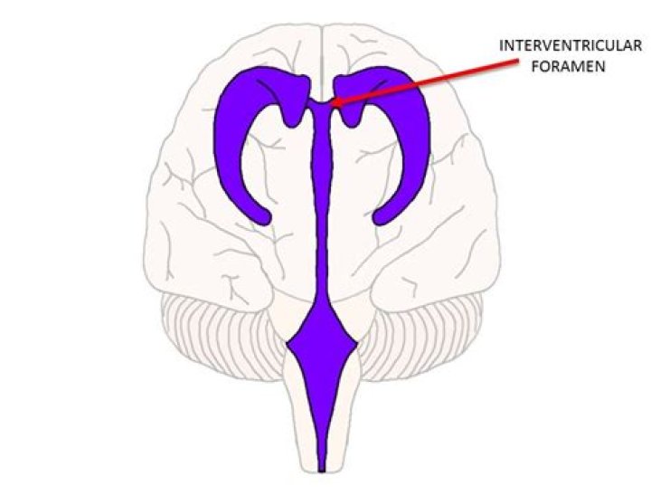
The interventricular foramen, also known as foramen of Monro, is part of the ventricular system and the connection between the third ventricle and the lateral ventricle.
What does interventricular foramen mean?
Interventricular foramen: An opening between the lateral and third ventricles in the system of four communicating cavities within the brain that are continuous with the central canal of the spinal cord.
Where is the interventricular foramen?
The interventricular foramen is located between the thalamus and anterior pillar of the fornix, at the anterior margin of the body. The 2 interventricular foramens (or foramina of Monro) connect the lateral ventricles with the third ventricle.What is the interventricular foramen heart?
A temporary opening between the developing ventricles of the heart. The ventricles arise as a single cavity is divided by the developing interventricular septum. Before the septum closes completely, the remaining opening between the two ventricles is termed the interventricular foramen.
Where is the 3rd ventricle?
The third ventricle is a narrow, funnel-shaped structure that lies in the center of the brain. It lies below the corpus callosum and body of the lateral ventricles, between the two thalami and walls of hypothalamus, and above the pituitary and midbrain (Fig. 28-1).
Are there two interventricular foramen?
The interventricular foramina are two holes (Latin: foramen, pl. foramina) that connect the left and the right lateral ventricles to the third ventricle. They are located on the underside near the midline of the lateral ventricles, and join the third ventricle where its roof meets its anterior surface.
What is the function of ventricles in the brain?
Aside from cerebrospinal fluid, your brain ventricles are hollow. Their sole function is to produce and secrete cerebrospinal fluid to protect and maintain your central nervous system.What does interventricular mean?
Definition of interventricular : situated or occurring between ventricles the interventricular septum of the heart interventricular brain hemorrhage.
What is atria and atrium?The upper two heart chambers are called atria. Atria are separated by an interatrial septum into the left atrium and the right atrium. The lower two chambers of the heart are called ventricles. Atria receive blood returning to the heart from the body and ventricles pump blood from the heart to the body.
Article first time published onIs the interventricular foramen a single structure or paired?
These paired foramina allow for the flow of cerebrospinal fluid between lateral ventricles and third ventricle, and effacement or blockage results in non-communicating obstructive hydrocephalus.
What is the lamina terminalis?
The lamina terminalis is a thin sheet of gray matter and pia mater that attaches to the upper surface of the chiasm and stretches upward to fill the interval between the optic chiasm and the rostrum of the corpus callosum.
What causes hydrocephalus?
Hydrocephalus is caused by an imbalance between how much cerebrospinal fluid is produced and how much is absorbed into the bloodstream. Cerebrospinal fluid is produced by tissues lining the ventricles of the brain.
How is interventricular foramen formed?
The ventricles arise as a single cavity that is divided by the developing interventricular septum. Before the septum closes completely, the remaining opening between the two ventricles is termed the interventricular foramen.
What closes interventricular foramen?
The interventricular foramen is usually completely closed by week seven. Closure is accomplished by growth of membraneous tissue derived from the endocardial cushions, the interventricular septum and from the conus ridges formed within the truncus and extending to the interventricular septum.
What is the interventricular septum composed of?
The interventricular septum is a complex structure composed of muscular and fibrous tissue.
How many ventricles does the brain have?
The cerebral ventricular system is made up of 4 ventricles that include 2 lateral ventricles (1 in each cerebral hemisphere), the third ventricle in the diencephalon, and the fourth ventricle in the hindbrain. Inferiorly, it is continuous with the central canal of the spinal cord.
Does the body produce spinal fluid?
The brain produces roughly 500 mL of cerebrospinal fluid per day, at a rate of about 25 mL an hour. This transcellular fluid is constantly reabsorbed, so that only 125–150 mL is present at any one time. CSF volume is higher on a mL per kg body weight basis in children compared to adults.
Which ventricle is located in the cerebrum?
They are located in the largest part of the brain, the cerebrum. The third ventricle is in the diencephalon, located in the center of the brain. The fourth ventricle is located in the hindbrain. Each lateral ventricle, one on each side of the brain, sits in a “C” shape.
What is the fourth ventricle of the brain?
The fourth ventricle contains cerebrospinal fluid. It has a diamond shape and is located in the upper portion of the medulla. … The main function of this ventricle is to protect the human brain from trauma (via a cushioning effect) and to help form the central canal, which runs the length of the spinal cord.
What is the hole in third ventricle?
The third ventricle is one of the four ventricles in the brain that communicate with one another. As with the other ventricles of the brain, it is filled with cerebrospinal fluid, which helps to protect the brain from injury and transport nutrients and waste.
What structure produces CSF?
CSF is produced mainly by a structure called the choroid plexus in the lateral, third and fourth ventricles. CSF flows from the lateral ventricle to the third ventricle through the interventricular foramen (also called the foramen of Monro).
What is the purpose of interventricular septum?
The interventricular septum divides the right and left ventricles, running in the plane of the anterior and posterior interventicular grooves. Septation of the ventricles occurs in the fetus within 7 weeks of gestation, achieved by the formation of this embryologically heterogenous structure 6.
What does epidural mean in medical terms?
noun. Definition of epidural (Entry 2 of 2) : an injection of a local anesthetic into the space outside the dura mater of the spinal cord in the lower back region to produce loss of sensation especially in the abdomen or pelvic region.
What does intercostal mean in medical terms?
Definition of intercostal : situated or extending between the ribs intercostal spaces intercostal muscles.
What does the third ventricle control?
The third ventricle can be described as a cuboid structure that has a roof, floor and four walls (anterior, posterior, and two lateral). Similar to the other brain ventricles, the main function of the third ventricle is to produce, secrete and convey cerebrospinal fluid.
What is brain stem & ventricles explain its functions?
brainstem, area at the base of the brain that lies between the deep structures of the cerebral hemispheres and the cervical spinal cord and that serves a critical role in regulating certain involuntary actions of the body, including heartbeat and breathing.
Which structures reabsorb CSF from the subarachnoid space?
The CSF from the subarachnoid space is eventually reabsorbed through outpouchings into the superior sagittal sinus (SSS) known as the arachnoid granulations. Arachnoid granulations act as an avenue for CSF reabsorption into the blood circulation through a pressure-dependent gradient.
What is the left auricle of the heart?
The left auricle, also known as the left atrial appendage (LAA), is a flap of heart wall on the anterior surface of the left atrium of the heart. … The name auricle comes from the Latin word auricula, which means “ear” and refers to the floppy dog-ear shape of the auricle.
What 3 vessels fill the right atrium?
The blood vessels include the superior and inferior vena cava. These bring blood from the body to the right atrium. Next is the pulmonary artery that carries blood from the right ventricle to the lungs.
What are venules?
Definition of venule : a small vein especially : any of the minute veins connecting the capillaries with the larger systemic veins.