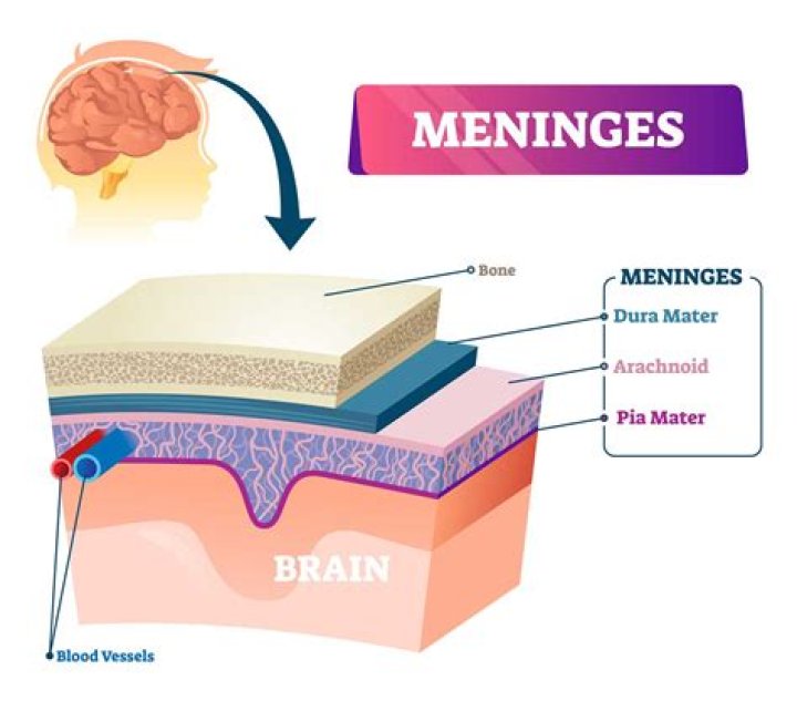
Meninges are formed by three tissue membranes that are primarily known as wrappers of the brain. They consist of dura mater, arachnoid and pia mater. The dura mater or pachymeninx (pachy-thick) is the outer membrane and forms a sac that envelops the other meningeal layers.
Are meninges epithelial?
Segregating the outer and inner meningeal compartments is the epithelial-like arachnoid barrier cells, connected by tight and adherens junctions, which regulate the movement of pathogens, molecules, and cells into and out of the cerebral spinal fluid (CSF) and brain parenchyma.
What are the 3 membranes of the meninges?
Three layers of membranes known as meninges protect the brain and spinal cord. The delicate inner layer is the pia mater. The middle layer is the arachnoid, a web-like structure filled with fluid that cushions the brain. The tough outer layer is called the dura mater.
Are meninges connective tissue membrane?
The meninges are the connective tissue coverings of the brain and spinal cord. The outermost layer is the dura mater, a dense and tough tissue that is reduplicated to form the periosteum of the inner skull. Beneath the dura is the arachnoid, a network of loose connective tissue that lacks blood vessels.What is the cerebral membrane?
The cerebral cortex, also known as the cerebral mantle, is the outer layer of neural tissue of the cerebrum of the brain in humans and other mammals. The cerebral cortex mostly consists of the six-layered neocortex, with just 10% consisting of allocortex.
What is between the arachnoid and pia mater?
The subarachnoid space is the cerebrospinal fluid-filled space that exists between the arachnoid and the pia.
Is the meninges dorsal or ventral?
The meninges (dura mater, arachnoid membrane, and pia mater) enclose the spinal cord and on the left separately ensheathe dorsal and ventral roots to the level of the spinal nerve, where meninges blend with connective tissue surrounding spinal nerves. On the right only the pia mater ensheathes the spinal roots.
What is the order of the membrane layer in the meninges?
The membrane layers (discussed in detail below) from the outside in are the: dura mater, arachnoid mater, and pia mater.Is the most superficial membrane of the meninges?
The most superficial layer of the meninges is the dura mater.
Is Pons part of midbrain?The pons (Latin for “bridge”) is part of the brainstem that in humans and other bipeds lies inferior to the midbrain, superior to the medulla oblongata and anterior to the cerebellum.
Article first time published onWhat are meninges made of?
The meninges are three protective membrane layers surrounding the brain and spinal cord. They are composed of the pia (closest to the CNS), arachnoid, and dura (outermost layer), and contain blood vessels and enclose the cerebrospinal fluid.
What is the arachnoid villi?
Arachnoid granulations or villi are growths of arachnoid membrane into the dural sinuses, through which the CSF enters the venous system from the subarachnoid space. 1. Arachnoid villi are microscopic, whereas arachnoid granulations represent distended villi and are visible to the naked eye.
What is spiral cord?
A column of nerve tissue that runs from the base of the skull down the center of the back. It is covered by three thin layers of protective tissue called membranes. The spinal cord and membranes are surrounded by the vertebrae (back bones).
What are the spinal meninges?
INVESTING MEMBRANES (MENINGES) Three membranes surround the spinal cord: The outermost is the dura mater (dura), the next is the arachnoid, and the innermost is the pia mater (pia) (Figs 6–1 and 6–2). The dura is also called the pachymeninx, and the arachnoid and pia are called the leptomeninges.
What do you mean by meninges?
Listen to pronunciation. (meh-NIN-jeez) The three thin layers of tissue that cover and protect the brain and spinal cord.
What do the spinal meninges do quizlet?
Provide physical stability and shock absorption while providing oxygen and nutrient to the spinal cord tissue.
Where are meninges located?
Brain meninges are three-layer tissue envelopes that have a protective, supportive and metabolic role. They are located between the brain and the skull and between the spinal cord and spinal vertebrae and are constructed of loose and dense connective tissues.
Where do the meninges attach?
The dura mater is attached to the skull, whereas in the spinal cord, the dura mater is separated from the vertebrae by a space called the epidural space, which contains fat and blood vessels. The arachnoid is attached to the dura mater, while the pia mater is attached to the central nervous system tissue.
What are the meninges quizlet?
Protect the brain from injury and is made up of three layers: dura mater, arachnoid mater, and pia mater. … Dura mater. Two layers: superficial Periosteal Layer attachs to the skull and deeper Meningeal Layer forms the true external covering of the brain.
Are the meninges part of the central nervous system?
Central nervous system: The central nervous system (2) is a combination of the brain (1) and the spinal cord (3). The CNS is covered with three layers of protective coverings called meninges (from the Greek word for membrane). The outermost layer is the dura mater (Latin for “hard mother”).
Where is the origin of the meninges?
The cranial meninges originate from a mesenchymal sheath on the surface of the developing brain, called primary meninx, and undergo differentiation into three layers with distinct histological characteristics: the dura mater, the arachnoid mater, and the pia mater.
What is the role of the meninges?
The primary function commonly attributed to meninges and CSF is to protect the central nervous system (CNS). This is mainly because meninges provide a tight anchoring of the CNS to the surrounding bones able to prevent side-to-side movement and providing stability.
How are meninges linked to each other?
The two dural layers are firmly attached to each other, except in places where they separate to enclose the dural venous sinuses. In these places, the meningeal layer projects inward, towards the cerebral tissue, forming the fibrous septa that partially separate the cranial cavity.
What nerve innervates the diaphragm quizlet?
Terms in this set (5) phrenic nerve arises from this plexus and innervates the diaphragm. The phrenic nerve arises from the 3rd, 4th and 5th cervical spinal nerves.
What is meninges in biology?
The meninges are membranous layers surrounding the central nervous system. In the head, the meninges lie between the brain and the skull, and interact closely with both during development.
Where is white matter?
White matter is found in the deeper tissues of the brain (subcortical). It contains nerve fibers (axons), which are extensions of nerve cells (neurons). Many of these nerve fibers are surrounded by a type of sheath or covering called myelin. Myelin gives the white matter its color.
What are the 4 ventricles of the brain?
The fourth ventricle is one of the four connected fluid-filled cavities within the human brain. These cavities, known collectively as the ventricular system, consist of the left and right lateral ventricles, the third ventricle, and the fourth ventricle.
Is the thalamus part of the midbrain?
The thalamus is a paired gray matter structure of the diencephalon located near the center of the brain. It is above the midbrain or mesencephalon, allowing for nerve fiber connections to the cerebral cortex in all directions — each thalamus connects to the other via the interthalamic adhesion.
What is posterior to the midbrain?
The posterior surface of the midbrain is called the tectum, or roof, of the midbrain. The tectum features four tubercles on its surface which lie inferior to the pineal gland.
Is the medulla in the midbrain?
BrainstemPartsMedulla, Pons, MidbrainIdentifiersLatintruncus encephaliMeSHD001933
What is the midbrain?
The midbrain is the topmost part of the brainstem, the connection central between the brain and the spinal cord. There are three main parts of the midbrain – the colliculi, the tegmentum, and the cerebral peduncles.