The neurovascular bundle consists of, from above downwards, intercostal vein, artery and nerve. See Clinical box 3.2. The intercostal nerves are the anterior rami of the first 11 thoracic nerves. These supply the intercostal muscles, the skin of the chest wall as well as the parietal pleura.
Which muscles do the intercostal nerves supply?
Branches. Numerous slender muscular filaments supply the Intercostales, the Subcostales, the Levatores costarum, the Serratus posterior superior, and the Transversus thoracis. At the front of the thorax some of these branches cross the costal cartilages from one intercostal space to another.
Where in the intercostal space does the neurovascular bundle run?
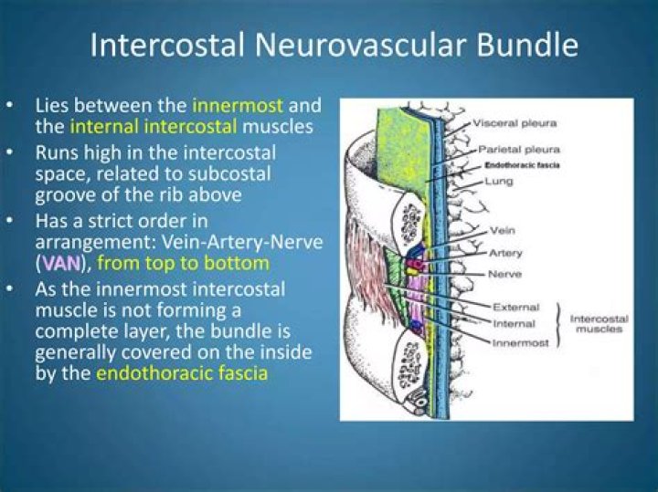
Neurovascular bundle. The neurovascular bundle, located in the costal groove in the undersurface of each rib, between the internal intercostal muscle and innermost intercostal muscle, supplies much of the innervation and vascular supply to the thoracic wall.
What does the intercostal supply?
The posterior intercostal arteries are branches of the superior intercostal artery (upper two spaces) and the descending aorta (lower nine spaces). They supply the chest wall, parietal pleura, and, through their dorsal branches, the skin and muscles of the back and the spine and its contents.Which is the highest structure in neurovascular bundle of 1st intercostal space?
Neurovascular bundles The neurovascular bundle has a strict order of vein-artery-nerve (VAN), from top to bottom. This neurovascular bundle runs high in the intercostal space, and the smaller collateral neurovascular bundle runs just superior to the lower rib of the space (in the order NAV from superior to inferior).
What does the Subcostal nerve innervate?
Subcostal nerve (T12). The subcostal nerve provides sensory innervation to the region under the umbilicus and also provides motor innervation to the pyramidalis and quadratus lumborum muscles.
Which main structure do the intercostal nerves supply quizlet?
Found within the costal grooves on the inferior side of each rib along with a vein and an artery. Gives motor innervation to intercostal muscles and brings sensory information from anterior and lateral chest and abdominal wall. Thoracic spinal nerves supply deep muscles and skin of the back.
What does the posterior intercostal vein supply?
The posterior intercostal veins are veins that drain the intercostal spaces posteriorly. They run with their corresponding posterior intercostal artery on the underside of the rib, the vein superior to the artery. Each vein also gives off a dorsal branch that drains blood from the muscles of the back.What supplies anterior intercostal arteries?
The musculophrenic artery supplies the anterior intercostal arteries of spaces 7 to 9 after it branches off the internal thoracic.
What travels in the costal groove?The posterior intercostal artery travels in the costal groove between the intercostal vein and nerve. The vessel first passes in the endothoracic fascia before passing between the internal and innermost intercostal muscles.
Article first time published onWhat is a neurovascular structure?
A neurovascular bundle is a structure that binds nerves and veins (and in some cases arteries and lymphatics) with connective tissue so that they travel in tandem through the body.
Where are intercostal spaces?
The intercostal spaces, also known as interspaces, are the space between the ribs. There are 11 spaces on each side and they are numbered according to the rib which is the superior border of the space.
How do you find the intercostal space?
From the angle of Louis, move your fingers to the right and you will feel a gap between the ribs. This gap is the 2nd Intercostal space. From this position, run your fingers downward across the next rib, and the next one. The space you are in is the 4th intercostal space.
Where is 4th intercostal space?
The location of the 4th and 5th intercostal space is related to the length of the sternum. It is 77% of the sternal length that measures 15cm for the 4th intercostal space. The position of the V1 and V2 electrodes decreases to 57% when the sternal length is 26cm.
What does the Musculophrenic artery supply?
The musculophrenic artery runs along the costal slips of the diaphragm. It supplies the 7th, 8th and 9th intercostal spaces with paired anterior intercostal arteries, as well as fine branches that supply the superior part of the anterior abdominal wall.
Where is 5th intercostal space?
The apex (the most inferior, anterior, and lateral part as the heart lies in situ) is located on the midclavicular line, in the fifth intercostal space. It is formed by the left ventricle. The base of the heart, the posterior part, is formed by both atria, but mainly the left.
How intercostal nerve is formed?
The intercostal nerves arise from the anterior rami of the thoracic spinal nerves from T1 to T11. The anterior division of the twelfth thoracic nerve is not technically grouped with the other intercostal nerves as it enters the abdominal wall; this nerve is instead referred to as the subcostal nerve.
Which branch of a spinal nerve serves the deep muscles and skin?
Posterior rami carry visceral motor, somatic motor, and sensory information to and from the skin and deep muscles of the back. The posterior rami remain distinct from each other, and each innervates a narrow strip of skin and muscle along the back, more or less at the level from which the ramus leaves the spinal nerve.
Where is the Subcostal nerve located quizlet?
**Subcostal nerves are found underneath the floating ribs, rather than in between true ribs.
Which structure is subcostal nerve related?
The subcostal nerve (anterior division of the twelfth thoracic nerve) is larger than the others. It runs along the lower border of the twelfth rib, often gives a communicating branch to the first lumbar nerve, and passes under the lateral lumbocostal arch.
What is innervated by the obturator nerve?
The anterior branch of the obturator nerve innervates the adductor longus, adductor brevis, and gracilis muscles, as well as giving innervation to the hip joint. … The obturator nerve originates from posterior divisions of L2, L3, and L4 spinal roots.
What are the thoracoabdominal nerves?
The thoracoabdominal nerves are derived from T7-T11 and form the inferior intercostal nerves. These nerves run along the internal obliques and the transversalis muscles. They then enter subcutaneous tissue to become the anterior cutaneous branches of the skin in the anterior abdominal wall.
What supplies the posterior intercostal arteries?
Intercostal arteriesSuppliesIntercostal muscles and intercostal spaceIdentifiersLatinArteriae intercostalesAnatomical terminology
What does the posterior intercostal artery come from?
The intercostal spaces are supplied by pairs of anterior and posterior intercostal arteries. The posterior intercostal arteries arise from the aorta and in part supply the spine and spinal cord and thus are considered segmental arteries.
What supplies the posterior chest wall?
Three arteries supply each intercostal space; the posterior intercostal artery and two branches of anterior intercostal arteries. These intercostal blood vessels run along with the nerves between the internal intercostal muscle and innermost intercostal muscles in the costal groove.
What artery supplies the thoracic wall?
The internal thoracic artery, aka the internal mammary artery, supplies the breast and the anterior chest wall. The internal thoracic artery travels along the inner surface of the anterior chest wall on both sides.
What do the intercostal veins do?
The intercostal veins are a group of veins which drain the area between the ribs (“costae”), called the intercostal space.
Where are the intercostal artery vein and nerve found?
The intercostal artery, vein, and nerve run along the inferior aspect of each rib, occasionally running underneath a ledge in the costal groove.
What is the function of the costal groove?
The costal groove in the inferior margin of each rib carries blood vessels and a nerve. Anteriorly, each rib ends in a costal cartilage. True ribs (1–7) attach directly to the sternum via their costal cartilage.
Which components form neurovascular bundle of the neck?
- the common carotid artery.
- parts of the internal carotid artery and the external carotid artery.
- the internal jugular vein.
- the vagus nerve.
- part of the recurrent laryngeal nerve.
- the deep cervical lymph nodes.
Which Plexus Innervates muscles of the neck?
The cervical plexus, composed of the anterior rami of C1 to C4 cervical roots, innervates most neck muscles and provides sensory innervation to the anterior and lateral neck.