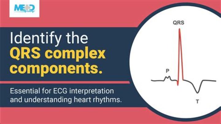
During the P Wave, the electrical impulses progress from the SA node through the intermodal atrial conduction tracts and most of the AV node. The DIRECTION of the P Wave in lead II is positive (upright).
Why are some ECG leads upside down?
It is normally upside down in VR and V1. If it is upside down in any other lead, then the likely causes are ischaemia or ventricular hypertrophy (Fig. 1.12).
Why the T wave is a positive upward deflection?
T and U waves The T wave represents ventricular repolarization. Generally, the T wave exhibits a positive deflection. The reason for this is that the last cells to depolarize in the ventricles are the first to repolarize.
What does tall QRS complex mean?
Tall QRS complexes are usually caused by hypertrophy of one or both ventricles, or by an abnormal pacemaker or aberrantly conducted beat. • Low voltage or abnormally small QRS complexes may be seen in obese patients, hyperthyroid patients and pleural effusion.What do high peaks on an EKG mean?
Your ECG Results The distance between these spikes shows your heart rate. If the distances are too short, too long, or irregular, it can be a sign of a problem. For example, spikes that are too close together are a sign of a rapid heartbeat or tachycardia. Each heartbeat will be made up of several spikes in activity.
What causes narrow complex tachycardia?
It can occur at any age, but there is an increased likelihood if the atria are diseased (hypertension, pulmonary disease, previous cardiac surgery, etc). The causes of atrial tachycardia include digoxin toxicity, coronary heart disease, cardiomyopathy, rheumatic heart disease and sick sinus syndrome.
What does a wavy EKG mean?
What do the wavy lines on my EKG readings mean? Electrical activity from the heart’s contraction creates the wavy lines on your EKG.
What is the treatment for wide complex tachycardia?
SVT will typically be managed with adenosine, Afib with WPWS will be treated with amiodarone, and Afib with aberrancy with either diltiazem or a beta-blocker. Typically, amiodarone will be the first-line drug of choice for all ventricular arrhythmias (VT, polymorphic VT, Vfib, etc.)What is Brugada syndrome?
Brugada (brew-GAH-dah) syndrome is a rare, but potentially life-threatening heart rhythm disorder that is sometimes inherited. People with Brugada syndrome have an increased risk of having irregular heart rhythms beginning in the lower chambers of the heart (ventricles).
Should I worry about inverted T waves?Inverted T-waves are not uncommon, and you don’t need to be overly anxious about them as long as you continue to feel well and have normal echocardiograms and stress tests.
Article first time published onWhat do T wave inversions mean?
Deep T wave inversions, as described previously, usually occur during the evolving phase of a Q wave MI (see Fig. 8-4B) and also sometimes with a non–Q wave MI (see Fig. 9-7). These deep inversions are the result of a delay in regional repolarization produced by the ischemic injury.
Can anxiety cause inverted T waves?
(HealthDay)—Depression and anxiety are independently, yet oppositely, associated with electrocardiographic (ECG) T-wave inversions, according to a study published in the Dec. 15 issue of The American Journal of Cardiology.
What does sinus tachycardia look like on an ECG?
Sinus tachycardia is recognized on an ECG with a normal upright P wave in lead II preceding every QRS complex. This indicates that the pacemaker is coming from the sinus node and not elsewhere in the atria, with an atrial rate of greater than 100 beats per minute.
Is sinus tachycardia a disease?
Sinus tachycardia is a normal response to physical exercise, when the heart rate increases to meet the body’s higher demand for energy and oxygen, but sinus tachycardia can also indicate a health problem. Thus, sinus tachycardia is a medical finding that can be either physiological or pathological.
Can anxiety cause an abnormal EKG?
Premature ventricular contractions is one of the manifestations of sympathetic over activity due to anxiety. However, anxiety might induce electrocardiographic (ECG) changes in normal person with normal heart, as in this documented case.
What wave is usually never seen on an EKG?
Strictly speaking, the atria contract a split second after the P wave begins. Because it is so small, atrial repolarization is usually not visible on ECG. In most cases, the P wave will be smooth and rounded, no more than 2.5 mm tall, and no more than 0.11 seconds in duration.
What is complex tachycardia?
A wide complex tachycardia (WCT) is simple enough to define: a cardiac rhythm with a rate >100 beats per minute and a QRS width >120 milliseconds (ms).
Is AFIB a narrow complex tachycardia?
Narrow complex tachycardias are Supraventricular tachycardias, meaning only that they originate above the ventricles. Rhythm: Irregular: atrial fibrillation, flutter with variable conduction, MAT.
Is narrow complex tachycardia serious?
Narrow QRS complex tachycardias are fast cardiac rhythms (generally more than 100 beats/min) with a QRS duration of 100 ms or less. Differential diagnosis of tachycardia with narrow QRS complex (shorter than 0.12 second). Although rarely life-threatening, these arrhythmias are a common source of morbidity.
What does Brugada look like on EKG?
Brugada syndrome is a disorder characterized by sudden death associated with one of several electrocardiographic (ECG) patterns characterized by incomplete right bundle-branch block and ST elevations in the anterior precordial leads.
What is a rare heart condition?
Restrictive Cardiomyopathy. Restrictive cardiomyopathy is the rarest form of heart-muscle disease.
What causes Romano Ward syndrome on the cellular level?
Mutations in the KCNQ1, KCNH2, and SCN5A genes are the most common causes of Romano-Ward syndrome. These genes provide instructions for making proteins that form channels across the cell membrane. These channels transport positively charged atoms (ions), such as potassium and sodium, into and out of cells.
What are 2 common cause of wide complex tachycardia?
Most commonly, wide-complex tachycardia or ventricular tachycardia originates from coronary artery disease. The presence of cardiomyopathies with or without left ventricular dysfunction are often already known.
Is wide complex tachycardia fatal?
Despite hemodynamic stability in some patients with ventricular tachycardia, incorrect or untimely diagnosis can be dangerous, if not fatal.
At what heart rate does tachycardia usually become symptomatic?
Heart rate greater than 150 bpm may be symptomatic; the higher the rate, the more likely the symptoms are due to the tachycardia.
Can high blood pressure cause an abnormal EKG?
High blood pressure Other aspects of heart disease may lead to an abnormal EKG. For example, people with high blood pressure are more likely to have an abnormal EKG reading.
What causes ST and T wave abnormality?
Factors affecting the ST-T and U wave configuration include: Intrinsic myocardial disease (e.g., myocarditis, ischemia, infarction, infiltrative or myopathic processes) Drugs (e.g., digoxin, quinidine, tricyclics, and many others) Electrolyte abnormalities of potassium, magnesium, calcium.
Can inverted T wave reversed?
In a recent review, reversible or permanent inverted T-waves were found in 38% of patients with congenital coronary artery-ventricular multiple micro-fistulas (MMFs)[27]. Hence, congenital MMFs may be included in the differential diagnosis of anterior chest wall T-wave inversion.
What causes Myocardial ischemia?
Causes of myocardial ischemia Myocardial ischemia occurs when blood flow to the heart muscle (myocardium) is obstructed by a partial or complete blockage of a coronary artery by a buildup of plaques (atherosclerosis). If the plaques rupture, you can have a heart attack (myocardial infarction).
What does anxiety look like on ECG?
The ECG changes in anxiety are: ST flattening, the commonest finding. Frank ST depression; not rare, especially in hyperventilation. T wave inversion.
Can EKG be wrong?
The study of 500 patients found a false positive reading between 77 and 82 percent in patients screened by electrocardiogram, and a false negative reading between 6 percent to 7 percent in the same patient population.