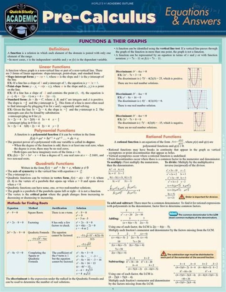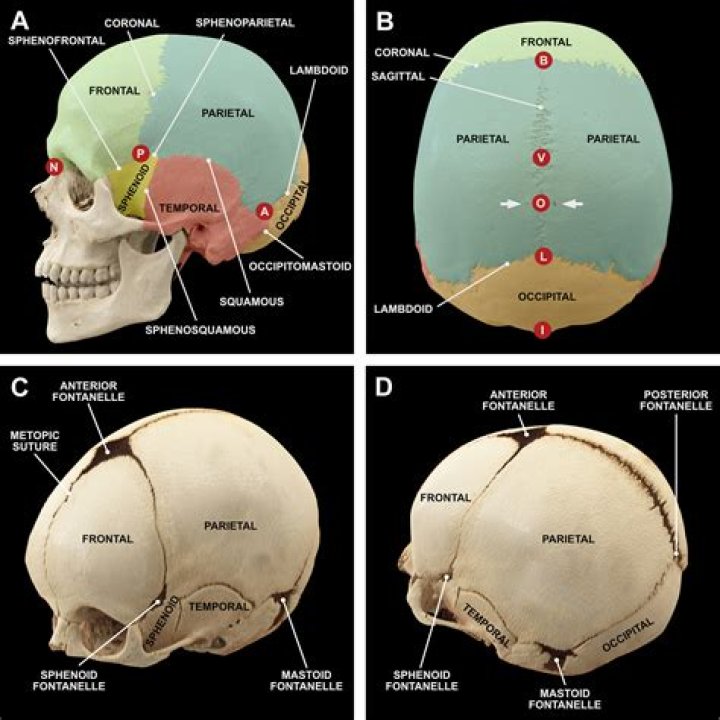
The calvaria is the top part of the skull. It is the upper part of the neurocranium
What does the medical term Calvarium mean?
Definition of calvarium : the portion of a skull including the braincase and excluding the lower jaw or lower jaw and facial portion.
What is another name for the Calvarium?
the dome of the skull.
Where is the Calvarium in the brain?
The calvaria definition is a simple one — the calvaria is the topmost part of the neural cranium, which protects the cranial cavity that houses the brain.What is the parietal part of the skull?
parietal bone, cranial bone forming part of the side and top of the head. In front each parietal bone adjoins the frontal bone; in back, the occipital bone; and below, the temporal and sphenoid bones. The parietal bones are marked internally by meningeal blood vessels and externally by the temporal muscles.
What is a Calvarial mass?
Calvarial lesions may be benign or malignant; fortunately, benign tumours are the most commonly encountered lesions [1–6]. The skull vault is formed by the frontal, parietal, temporal and occipital bones and parts of the zygoma and sphenoid bone.
Where is the right parietal calvarium?
Anatomical terms of bone The calvaria is the top part of the skull. It is the upper part of the neurocranium and covers the cranial cavity containing the brain. It forms the main component of the skull roof. The calvaria is made up of the superior portions of the frontal bone, occipital bone, and parietal bones.
What is Calvarial defect?
Definition. A localized defect in the bone of the skull resulting from abnormal embryological development. The defect is covered by normal skin. In some cases, skull x-rays have shown underlying lytic bone lesions which have closed before the age of one year. [ from HPO]What is boundary between base and calvarium?
The calvaria or norma verticalis is the outline of the skull as viewed from above. The border between the calvaria and the skull base passes through the squama occipitalis, angulus mastoideus ossis parietalis, pars squamosa ossis temporalis, ala major ossis sphenoidalis, and squama frontalis.
What is a Calvarial lesion?Calvarial lesions can originate within the calvarium (primary lesions), or invade the calvarium from the scalp or meninges. The calvarium is composed of a cortical outer table, marrow space (diploë), and a cortical inner table.
Article first time published onWhere is Glabella?
Your “glabella” is the skin on your forehead, between your eyebrows and above your nose. When you make facial expressions, that skin is moved by the muscles on your forehead.
What is the Forum Magnum?
The foramen magnum (Latin: great hole) is a large, oval-shaped opening in the occipital bone of the skull. It is one of the several oval or circular openings (foramina) in the base of the skull. … It also transmits the accessory nerve into the skull. The foramen magnum is a very important feature in bipedal mammals.
Is it easy to remove the calvaria?
A slender chisel is now driven into the notch made by the saw and the calvarium is removed by a gentle prying force. This procedure is readily carried out with a hand or electric saw. Visualization of the cranial cavity and access to its contents are entirely adequate.
Why is it called parietal bone?
The parietal bones (/pəˈraɪ. ɪtəl/) are two bones in the skull which, when joined together at a fibrous joint, form the sides and roof of the cranium. In humans, each bone is roughly quadrilateral in form, and has two surfaces, four borders, and four angles. It is named from the Latin paries (-ietis), wall.
What parts of the brain are in the parietal lobe?
Parietal lobePart ofCerebrumArteryAnterior cerebral Middle cerebralVeinSuperior sagittal sinusIdentifiers
What is the right parietal lobe responsible for?
The parietal lobes contain the primary sensory cortex which controls sensation (touch, pressure). Behind the primary sensory cortex is a large association area that controls fine sensation (judgment of texture, weight, size, and shape).
What is the main function of the parietal bones?
The function of the cranium, and hence the parietal bones, is to protect the underlying fragile brain. The parietal bone is slightly curved and has a quadrilateral shape. It has two surfaces, four borders and four angles. The borders articulate with the neighbouring skull bones to form various cranial sutures.
Is parietal bone strong?
(1700–5770). The average tensile strength of parietal compacta is less than that of compacta from long bones but the compressive strength, loaded both lengthwise and crosswise, is similar to that of compacta of other bones.
What is bony Calvarium intact?
“The bony calvaria is intact.” The calvaria is comprised of the upper frontal, temporal, parietal and occipital bones. … Just say “The calvaria is intact.” The skull is the skeleton of the head excluding the mandible. It is composed of the cranium and face.
Can bone islands be cancerous?
Most bone lesions are benign, not life-threatening, and will not spread to other parts of the body. Some bone lesions, however, are malignant, which means they are cancerous. These bone lesions can sometimes metastasize, which is when the cancer cells spread to other parts of the body.
What is the meaning of osteoma?
Osteomas are benign head tumors made of bone. They’re usually found in the head or skull, but they can also be found in the neck. While osteomas are not cancerous, they can sometimes cause headaches, sinus infections, hearing issues or vision problems – however, many benign osteomas don’t require treatment at all.
What is skull base?
The skull base consists of several bones that form the bottom of the head and the bony ridge behind the eyes and nose. Many different kinds of tumors can grow in this area.
Where is sphenoid?
The sphenoid is an unpaired bone. It sits anteriorly in the cranium, and contributes to the middle cranial fossa, the lateral wall of the skull, and the floor and sides of both orbits. It has articulations with twelve other bones: Unpaired bones – Occipital, vomer, ethmoid and frontal bones.
Where is maxillary?
The maxilla is the bone that forms your upper jaw. The right and left halves of the maxilla are irregularly shaped bones that fuse together in the middle of the skull, below the nose, in an area known as the intermaxillary suture.
What is rat calvaria?
The rat calvarial defect can be used to evaluate bone regeneration and screen different biomaterials or tissue engineering constructs before moving to larger animals for potential translation to human applications in the craniofacial complex.
How many bones make up the calvaria?
The calvaria is composed of 5 bones: Frontal, parietal, occipital, temporal, and sphenoid (greater wings) bones that are primarily connected by the major sutures, including the coronal, sagittal, and lambdoid sutures. The metopic suture is variably seen in adults. There are many normal variants of the skull.
What are Calvarial metastases?
CALVARIAL metastases are found in 15%–25% of all. cancer patients, most often in those with systemic bony metastatic disease. Metastasis occurs via he- matogenous spread, retrograde seeding through Batson’s venous plexus, or direct extension through cranial foram- ina.
Are lucent lesions cancerous?
Lucent lesions of the sternum should be considered malignant until proven otherwise (Helms CA, personal communication, 1983). Keep in mind that the classic descriptions of bone tumors that you spend so much time studying are for untreated lesions.
What is intraosseous meningioma?
Intraosseous meningioma, also referred to as primary intraosseous meningioma, is a rare subtype of meningioma that accounts for less than 1% of all osseous tumors. They are the most common type of primary extradural meningiomas 6.
Why is glabella important?
The skin of the glabella may be used to measure skin turgor in suspected cases of dehydration by gently pinching and lifting it. When released, the glabella of a dehydrated patient tends to remain extended (“tented”), rather than returning to its normal shape.
Is glabella a muscle?
Understanding Glabellar Lines When you wrinkle your face by talking, yawning, laughing, or making expressions, the glabella is the skin most affected by the muscle activity beneath the surface.