The arrector pili muscle (APM) consists of a small band of smooth muscle that connects the hair follicle to the connective tissue of the basement membrane.
What are arrector pili muscles quizlet?
Definition of ARRECTOR PILI MUSCLE. : one of the small fan-shaped smooth muscles associated with the base of each hair that contract when the body surface is chilled and erect the hairs, compress an oil gland above each muscle, and produce the appearance of goose bumps—called also erector pili muscle, pilomotor muscle.
What is the function of the Arrector pili muscle quizlet?
The arrector pili muscles are small muscles attached to hair follicles in mammals. Contraction of these muscles causes the hairs to stand on end, known colloquially as goose bumps.
What is the histology of the Arrector pili muscle?
The arrector pili muscle is a small bundle of smooth muscle cells associated with the hair follicle. Contractions of this muscle elevate the hair, forming goose bumps, to release heat and help sebum to be released from gland into duct. This is a photo at low magnification of some hairs.Does the Arrector pili muscle has striations?
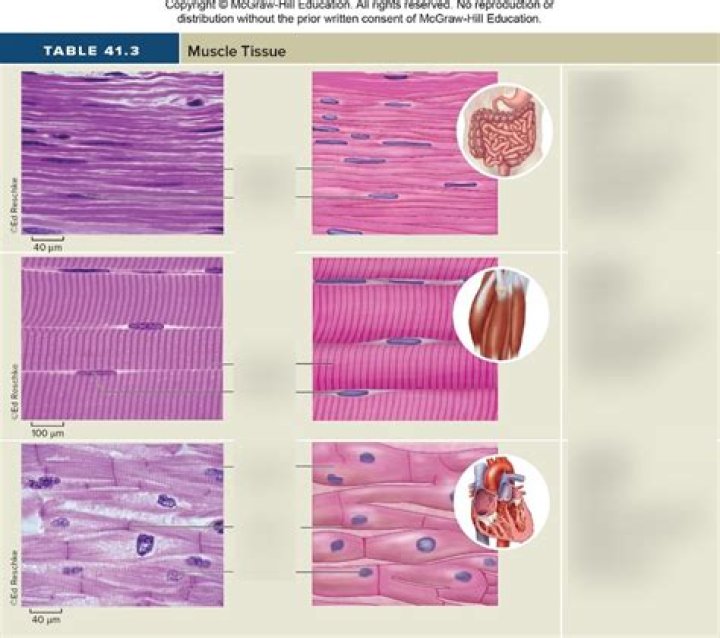
Perhaps because its action is most varied, striated muscle has been studied most extensively. This type of muscle is composed of numerous cylindrically shaped bundles of cells, each enclosed in a sheath called the sarcolemma.
Which type of tissue lines the follicles of the thyroid glands?
Thyroid follicular cells form a simple cuboidal epithelium and are arranged in spherical thyroid follicles surrounding a fluid filled space known as the colloid.
What type of tissue is the subcutaneous layer?
Subcutaneous tissue is the deepest layer of your skin. It’s made up mostly of fat cells and connective tissue. The majority of your body fat is stored here.
What type of tissue makes up the hair follicle?
Hair follicles (Fig. 6.3) are comprised of pockets of epithelium, which are continuous with the superficial epidermis and extend deep into the dermis. A hair follicle forms a bulb around the specialized dermal cells, the dermal papillae.What do dermal papillae do?
You should notice that the dermis extends up into the epidermis in structures called dermal papillae. These have two functions. First, they help adhesion between the dermal and epidermal layers. Second, in areas of thick skin like this, they provide a large surface area, to nourish the epidermal layer.
Which layer of cells in the epidermis contains melanocytes?The basal cell layer is also known as the stratum germinativum due to the fact that it is constantly germinating (producing) new cells. The basal cell layer contains cells called melanocytes.
Article first time published onWhat moves by contraction of the arrector pili muscles in the dermis?
The arrector pili muscles, also known as hair erector muscles, are small muscles attached to hair follicles in mammals. Contraction of these muscles causes the hairs to stand on end, known colloquially as goose bumps (piloerection).
Where can you find stratum lucidum?
Stratum lucidum, 2-3 cell layers, present in thicker skin found in the palms and soles, is a thin clear layer consisting of eleidin which is a transformation product of keratohyalin.
Where are apocrine sweat glands most abundant?
Apocrine glands develop in areas abundant in hair follicles, such as on your scalp, armpits and groin.
Which is a voluntary muscle tissue?
skeletal muscle, also called voluntary muscle, in vertebrates, most common of the three types of muscle in the body. Skeletal muscles are attached to bones by tendons, and they produce all the movements of body parts in relation to each other.
Is muscle subcutaneous tissue?
Subcutaneous tissue is the deepest skin layer that lies closest to the muscle. This layer has other names, including superficial fascia, hypodermis, subcutis, and tela subcutanea.
What is hair erector muscle?
Arrector Pili Muscle – This is a tiny muscle that attaches to the base of a hair follicle at one end and to dermal tissue on the other end. In order to generate heat when the body is cold, the arrector pili muscles contract all at once, causing the hair to “stand up straight” on the skin.
Is subcutaneous tissue considered soft tissue?
Soft tissue tumors can occur anywhere in the body, but if they occur subcutaneously, patients can easily notice a subcutaneous soft tissue mass. Therefore, it is possible to determine through recording, the growth speed of the mass, which is often difficult to obtain with deep-situated soft tissue masses.
What tissue is under skin?
Adipose tissue is present below the skin and between the internal organs. It stores fat. It functions as a shock absorber and insulating layer. ___ is the connective tissue beneath the skin that separates muscles and other internal organs.
Is fascia considered subcutaneous tissue?
According to standard textbooks of anatomy, the superficial fascia or “subcutaneous tissue” is described as a layer of loose areolar connective or adipose tissue that connects the skin to the underlying bones or deep fascia (3).
What is follicular epithelium?
The follicular epithelium originates as a few flattened cells derived from the germinal epithelium. Primary follicles are usually situated just under the tunica albuginea; secondary follicles lie deeper in the cortex.
What type of tissue lines the follicles of the thyroid glands quizlet?
Which type of tissue lines the follicles of the thyroid glands? glandular epithelium. A (An) gland does not branch before reaching the glandular cells or secretory part. A (An) gland branches repeatedly before reaching the glandular cells or secretory part.
What type of tissue is adipose tissue quizlet?
Type of connective tissue consisting mainly of fat cells (adipocytes), specialized to synthesize and contain large globules of fat, within a structural network of fibers.
Is dermis epithelial tissue?
Layers of Skin The skin is composed of two main layers: the epidermis, made of closely packed epithelial cells, and the dermis, made of dense, irregular connective tissue that houses blood vessels, hair follicles, sweat glands, and other structures.
What kind of epithelial tissue is skin?
Epidermis, the epithelial layer of skin, is primarily protective. This layer, consisting of keratinized stratified squamous epithelium, is tough, relatively impermeable, and self-replacing. These functional qualities are conferred by the epidermis’ principal cell type, the keratinocyte.
What type of epithelium is found in the epidermis quizlet?
The epidermis is the outermost layer of the skin and is composed of keratinized stratified squamous epithelium.
Is a hair follicle composed of epithelium and connective tissue?
The correct answer is True. A hair follicle is composed of both epithelial tissue and connective tissue.
Is hair epithelial tissue?
Hair is made in sac-like structures, called follicles. The follicles are rather simple organs that go through growth and rest phases. They are made of epithelial cells3, continuous with the surface epidermis (outermost skin layer) (Figure 4).
Is hair connective tissue?
Hair follicles originate in the epidermis and have many different parts. Hair is a keratinous filament growing out of the epidermis. … The hair bulb surrounds the hair papilla, which is made of connective tissue and contains blood capillaries and nerve endings from the dermis (Figure 1).
What is the outermost layer of the epidermis?
The outermost layer of the epidermis (stratum corneum) holds in water and keeps your skin hydrated and healthy. Producing new skin cells. New skin cells develop at the bottom layer of your epidermis (stratum basale) and travel up through the other layers as they get older.
Which layer of cells in the epidermis contains melanocytes quizlet?
Melanocytes are derived from neural crest cells and primarily produce melanin, which is responsible for the pigment of the skin. They are found between cells of stratum basale and produce melanin. UVB light stimulates melanin secretion which is protective against UV radiation, acting as a built-in sunscreen.
What type of cells are melanocytes?
Melanocytes. Melanocytes are dendritic, pigment-producing cells located in the basal cell layer (Figs 2.4, 2.5). They protect the skin from ultraviolet radiation. Individuals with little or no pigment develop marked sun damage and numerous skin cancers.